Once the molecule file is fully loaded, the image at right will become live. At that time the "activate 3-D" icon ![]() will disappear.
will disappear.
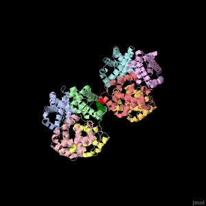
Simulation with and without the sickle cell mutation
In the previous slide, the sickle cell gene mutation that causes glutamic acid to be replaced with valine was shown. A simulation of two hemoglobin
proteins with and without this mutation have been run to show how the sickle cell mutated glutamate "sticks" the two hemoglobin tetramers together so they
can form long strands. The simulations were run for 175 ns (175*10^-9 s) - an exceedingly short time, but long enough to see the difference caused by the
mutation.
The button "With Sickle Cell Mutation" appears below. Click it to see how the valine in position 6 keeps the two hemoglobin tetramers together over the course of the simulation.
The button "With Sickle Cell Mutation" appears below. Click it to see how the valine in position 6 keeps the two hemoglobin tetramers together over the course of the simulation.
The button "Without Sickle Cell Mutation" appears below. Click it to see how the two hemoglobin tetramers drift apart without the mutation, i.e., in their
native state due to the random fluctuations always present at the molecular level.